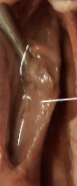
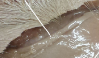
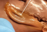
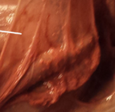
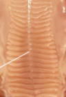
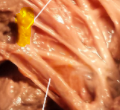
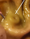
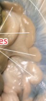
lab exam 2 digestive system
Primary tabs
45 cards submitted by Anonymous (not verified) on Wednesday, October 28th, 2020.
No Description Set
Bookmark to learn: Login to use bookmarks.
Edit this flashcard deck ...
Bookmark to learn: Login to use bookmarks.
Add to collection ... add lab exam 2 digestive system to your collections:
Help using Flashcards ...just like in real life ;)
- Try the tutorial for interactive instructions.
- Look at the card, do you know this one? Click to flip the card and check yourself.
- Mark card Right or Wrong, this card will be removed from the deck and your score kept.
- At any point you can Shuffle, Reveal cards and more via Deck controls.
- Continue to reveal the wrong cards until you have correctly answered the entire deck. Good job!
- Via the Actions button you can Shuffle, Unshuffle, Flip all Cards, Reset score, etc.
- Come back soon, we'll keep your score.“Repetition is the mother of all learning.”
- Signed in users can Create, Edit, Import, Export decks and more!.
Bookmark to learn: Login to use bookmarks.
Share via these services ...


Email this deck: Email this deck »
# Right & # Wrong of #